|
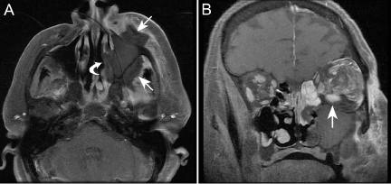
| Figure 1: MR imaging of a 58-year-old female (Case 1) with AFIFS. (A) Axial T1 MR with gadolinium and fat saturation demonstrates soft tissue swelling of the left cheek and opacification of the left maxillary sinus. Note, however, loss of contrast enhancement (LoCE) of the wall of the sinus, of the soft tissues anterior and posterior to the sinus (arrows), and of the nasal cavity lateral wall (curved arrow). LoCE of tissues is believed to correlate with tissue necrosis. (B) Coronal T1 MR with gadolinium and fat saturation shows extensive LoCE of the left maxilla and perimaxillary tissues, but also LoCE of the inflamed retrobulbar fat and periphery of the inferior rectus muscle (arrow). Normal extraocular muscles enhance on gadolinium-enhanced T1 MR. |