|
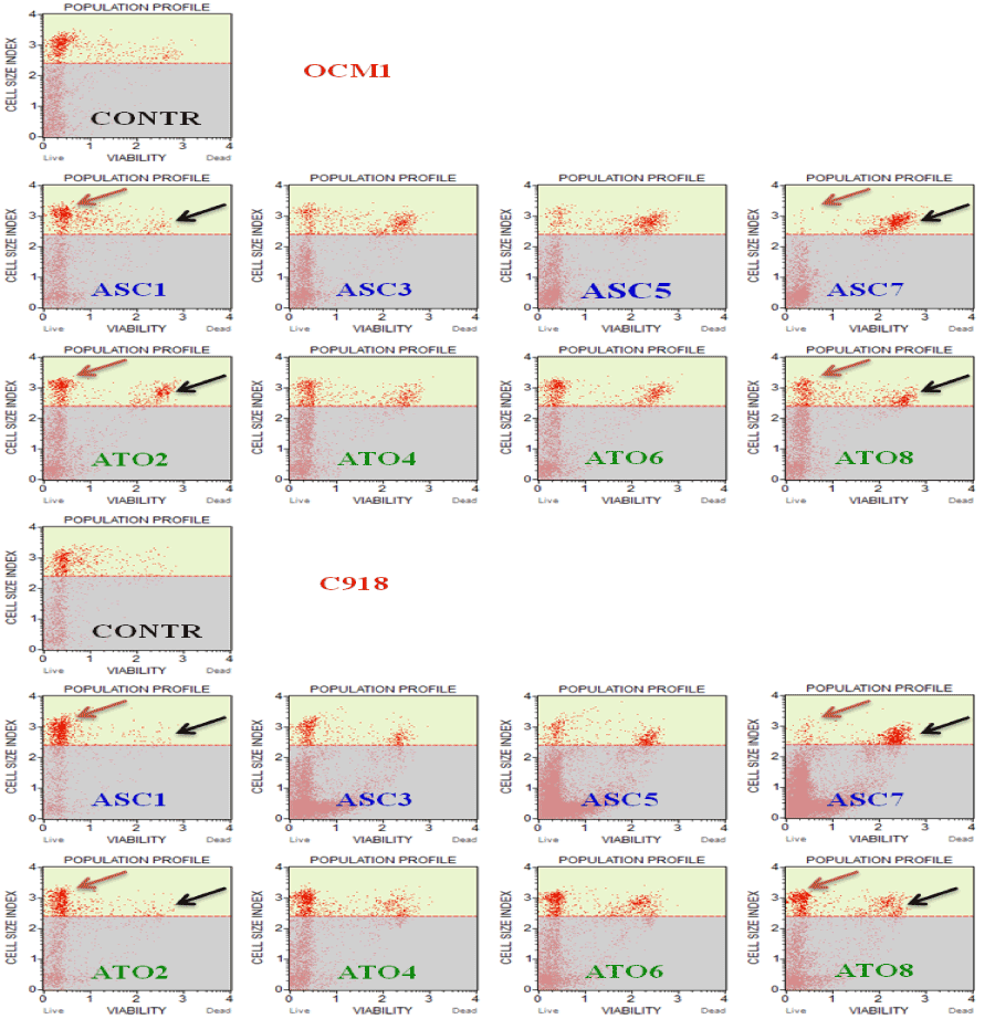
| Figure 2: Typical plot obtained with the Muse™ automated cell counter/analyzer (Merk - Millipore), by analyzing OCM1 and C918 UM cells after exposure to ASC 1, 3, 5, and 7 mM/ml, and ATO, 2, 4, 6, and 8 μg/ml. The red and black arrows in the plots indicate live and dead cells respectively. As shown in diagrams, a “cloud shift” can be appreciated, going from ASC1 (1 mM ASC) to ASC7 (7 mM ASC), for both OCM1 and C918 cell lines, thus indicating a progressive decrease of cell survival by increasing the concentration of ASC. The same does not apply to ATO. |