|
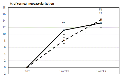
| Figure 1: Graphic rendering of the increasing corneal neovascularization in the lesioned eyes, showing that both fluorescence microscopy (continuous line) and capillaroscopy (dotted lines) give similar trends. **p<0.01 vs. start; ##p<0.01 vs. previous time point. |