 |
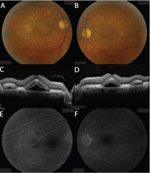
| Figure 2: Color fundus photos (A, B) of both eyes, demonstrating multiple yellowish, elevated, subretinal lesions in the macula of both eyes with subretinal fluid involving the fovea. Optical coherence tomography (C,D) of both eyes, showing multiple areas of subretinal fluid associated with decreased outer retinal band reflectivity and focal deposits of hyper-reflective material. Fluorescein angiography (E, F) of both eyes, revealing blockage without late leakage in the areas of the vitelliform lesions. |