|
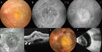
| Figure 3: Ophthalmic examination in case 3. A. Fundus photography at the initial visit. B. Early-phase fluorescein angiography. C. Late-phase fluorescein angiography. D. Optical coherence tomography shows bumpy areas of pigment epithelial detachment and an area of peaked pigment epithelial detachment from a flatter region of pigment epithelial detachment, raising suspicion for a polypoidal choroidal vasculopathy-associated polyp (arrow head). E. Fundus photography three months after the initial visit. F. B-scan ultrasonography one month after aflibercept injection. |