|
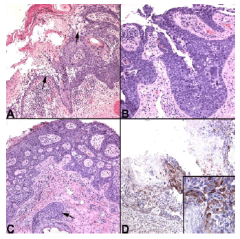
| Figure 2: (a) Photomicrograph of the enucleation specimen showing an area of extensive intraepithelial proliferation of cells with nuclear atypia and vacuolated cytoplasm (arrows) (H and E, 100X). (b) Some areas presented cells with high-grade nuclear atypia with scanty cytoplasm, simulating SqCC in situ (H and E, 400X). (c) Extension to the hair follicle was observed (arrow), explaining the madarosis of the patient (H and E, 200X). (d) Immunohistochemistry for BRST-1 showing positive stain on the neoplastic cells, especially in those well-differentiated sebaceous cells with vacuolated cytoplasm (BRST-1 100X). Inset, a closer view of the positive cells for BRST-1 (BRST-, 400X). |