|
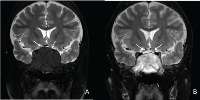
| Figure 3: MRI of the orbit and brain. (A) Coronal T2-weighted showing compression and stretching of bilateral prechiasmatic optic nerves (arrows). (B) Coronal T2-weighted showing reduction in the size of the tumor-like expansion along with improvement of compression and stretching of bilateral prechiasmatic optic nerves (arrows). |