|
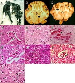
| Figure 1: Histopathological observations on the lesions of amprolium in the brains of the goat. A: Young lamb showing head retraction (opisthotonus). B and C: The distribution of the lesions was bilaterally symmetric foci of yellow malacic in the cerebral cortex. D and E: Perineuronal and perivascular oedema. F: Cerebral lesions were characterised by congestion. G: Increased perineuronal and perivascular spaces, neuronal degeneration with shrunken, angular/ triangular neurons, malacic foci. H: The lesions were comparatively mild and included vacuolation around neurons in grey matter, engorgement of blood vessels, perivascular and perineuronal clear zones indicative of edema. I and J: Polioencephalomalacia of the dorsal cerebral cortex with oedema and status spongiosus, edema and focal hemorrhage was detected in the white matter. |