 |
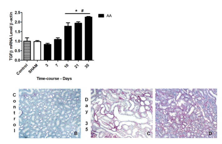
| Figure 2: Time course of fibrosis progression. Quantitative real-time PCR of TGF-ß mRNA was performed with kidney tissue from all experimental groups (A). Representative photographs of Sirius red staining in OSOM in control rat (B) and at day 35 (C&D) (x200). Values are means ± SEM. N=6 in each group. *p ≤ 0.05 versus SHAM rat at corresponding time-point. |