 |
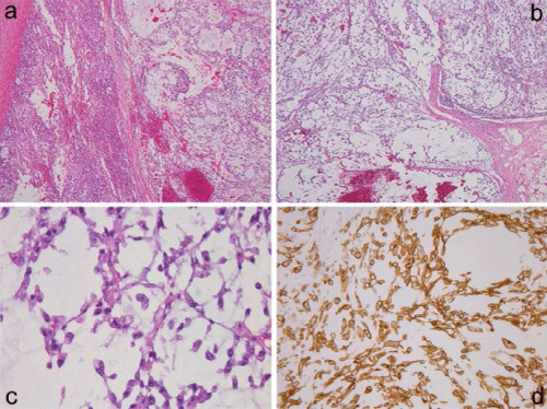
| Figure 2: Surgically removed right adrenal metastasis (a) showing transition between left-sided adenocarcinomatous components and right-sided markedly myxoid area. Low-power view of postmortem lymph node metastasis (b) composed of lobulated myxoid areas containing reticular cancerous proliferation, and the high-power view (c) showing polygonal or spindle cells arranged in short anastomosing cords with an abundant myxoid material, mimicking extraskeletal myxoid chondrosarcoma. Sarcomatous spindle cells in the liver metastasis showing cytokeratin 7 expression (d). |