 |
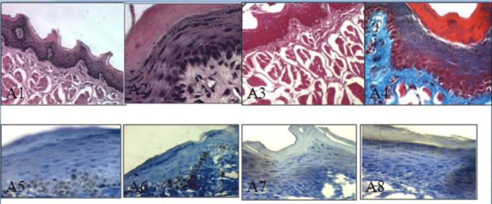
| Figure 3: A photomicrograph of buccal mucosa of rat in the control groups, revealed normal keratinized squamous epithelium with columinar basal cell layer and normal connective tissue (Al, H&E, x100; A2, H&E, x400), the muscles appear red (A3, PAS x100), ), and the collagen fibers appear blue in color in the connective tissue (A4, Trichrome x 400). Moderate Ki-67 immuno reactivity in nuclei of cells of basal and suprabasal cells layers in the first and second group respectively (AS, A6, immunohistochemistry x400), and mild cytoplasmic reaction to Bc1-2 in epithelial cells of the first and second group respectively (A7, A8, immunohistochemistry x 400). |