|
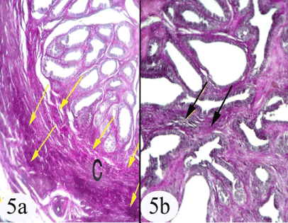
| Figure 5: a) A photomicrograph of the immature seminal gland showing the distribution of the elastic fibers in the capsule (arrow), capsule (C). b) Showing the distribution of elastic fibers in the inter-acinar connective tissue stroma (arrow). Stain: a,b) Weigert's elastic a) Obj. x4: Oc.x10 b) Obj. x10: Oc.x10 |