 |
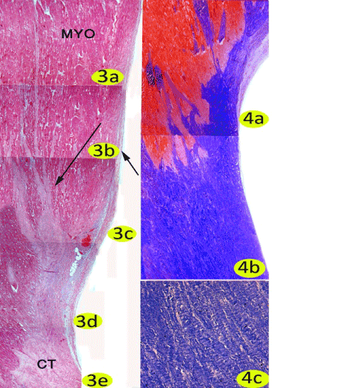
| Plate 2: Figure 3a, 3b, 3c, 3d, 3e: A photomicrograph of the Papillary muscle and the chordae tendinae showing the endothelium (short arrow), the myocardium (MYO), branches of chordae tendinae (long arrow) and the chordea tendinae (CT) Stain: all H& E all Obj.x5 : Oc.x10. (4a, 4b): showing the papillary muscle (red) and the chordea tendinae (blue) Stain: Azan all Obj.x5 : Oc.x10. (4c): High magnification of (Fig. 96) showing the collagen fibers in chordae tendinae. Stain: Azan Obj.x20 : Oc.x10. |