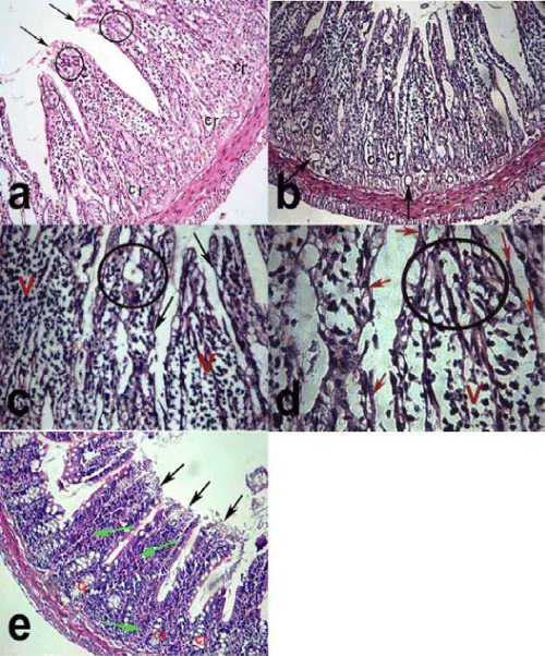
a) fusion of villi (circle), shedding of the surface epithelium (arrows) and distortion of the crypts (Cr). (H & E X100)
b) cystic dilatation of the crypts (Cr) at the bases of the villi and dilatation of the blood vessels (arrows). (H&E X100).
c) flattening of the villous surface epithelium (arrows) with loss of villus architecture (V) and fusion of villi (circle) especially near the apices. (H &E X200.)
d) flattened and shedded cells of the villous surface epithelium (arrows) with the loss of the villus architecture (V) and fusion of the villi (circle). (H &E X400)
e) showing distortion of the crypts (Cr) with blunting of the apices of the villi (black arrows) and intravillous hemorrhage (green arrows). (H & E X 100).