 |
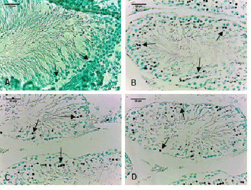
| Figure 3:Photomicrographs of TUNEL-stained testicular sections from controls (A) and rats exposed to 2 g/l of lead acetate (B, C, and D). Magnification 400x. Arrows indicated TUNEL-positive cells (spermatogonia; permatocytes I; spermatids) (A, B, C, D: Scale bar 20 μm) |