|
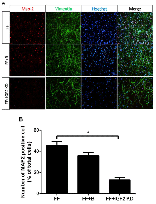
| Figure 2: Differentiation of RG-like cells into neurons after adding IGF2 knockdown virus. Few MAP2-positive neurons were detected in the FF+IGF2 KD group compared to the FF group (A). The MAP2-positive cells were quantified on statistics, and the results showed that the number of MAP2- positive cells in the FF+IGF2 KD group was lower than in the FF group (*P<0.05) (B). Scale bar, 50 μm. |