|
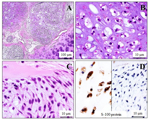
| Figure 2: Microscopic and immunohistochemical examination of the high-grade chondrosarcoma with spindle cell components arising from the cricoid cartilage. (A) Low-power view (H&E stains) showed that its multi-lobulated parts were composed of a solid proliferation of atypical chondrocyte-like cells, embedded in an abundant and basophilic chondroid matrix, very likely originated from the mature-looking cricoid cartilage (lower rt.). Bar=100 μm. (B) High-power view demonstrated that these neoplastic cells had a small to medium-sized and round shape, displaying hyperchromatic bi- or multi-nuclei, inconspicuous nucleoli, abundant clear vacuolated cytoplasm, and indistinct cellular borders (H&E stains). Bar=10 μm. (C) These neoplastic lobules coexisted with cellular spindled cell foci in part. Bar=10 μm. (D) In immunohistochemistry, the atypical chondrocyte-like cells were specifically positive for S-100 protein (lt.), but the atypical spindle cells were not (rt.) Bar=10 μm. |