|
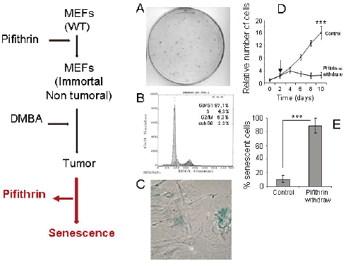
| Figure 3: Elimination of pifithrin-a from the media restoressenescence. Left) Schematic representation of the experiment. A)104 MEFs were seeded in a 10 cm plate and were treated with 10 Mpifithrin-a and 10 M DMBA. After 7 days pifithrin-a waseliminated from the media and cells grown for other 72 hrs inmedia without pifithrin-a. Then, the plate was fixed and A) stainedwith crystal violet to identify clones. B) processed to perform aFACS analysis of the proliferative status or C) analyze thephenotype of pifithrin-a- withdraw cells. D) Growth courve of cellstrated with pifithrin-a (control), or in which pifitrhin-a waswithdraw from the media. Arrow indicates the moment of thepifithin-a withdrawal. E) Cuantification of the percentage ofsenescent cells from 3 independent experiments. Senescent cellswere processed as indicated in materials and methods and morethan 150 cells counted in each experiment. Data shows the averageof senescent cells. Barrs show SD *: p<0.05, **: p<0.01, ***: p<0.001,Student’s T test. |