 |
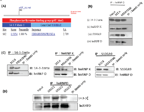
| Figure 3(A-D): Verification of hnRNPD binding partners in oral cancer. (A) 14-3-3 binding motif of hnRNPD. Photomicrograph showing presence of 14-3-3 binding motif, Mode 1 in hnRNPD polypeptide sequence as revealed by www.motif.scan.mit.edu. (B) Co-immunoprecipitation with hnRNPD in oral cancer cells. Western blot showing presence of (i) 14-3-3ζ; (ii) hnRNPK; (iii) S100A9 (iv) hnRNPD; proteins in immunoprecipitates of hnRNPD obtained from OSCC cells (SCC4 and MDA1986), while no band was seen in negative controls (i.e. beads only control). Input represents whole cell lysates from MDA1986 used as positive control for Western blotting. (C) Reverse immunoprecipitation verifying interaction partners of hnRNPD in oral cancer cells. Western blot showing presence of hnRNPD in immunoprecipitates of (i) 14-3-3ζ, (ii) hnRNP K; (iii) S100A9 in whole cell lysates obtained from OSCC cells (SCC4 and MDA1986), while no band was seen in negative controls (i.e. beads only control). Input represents whole cell lysates from MDA1986 used as positive control for Western blotting. (D) Confirmation of interaction of hnRNPD with 14-3-3ζ in OSCC tissue samples. Photomicrograph of a Western blot showing presence of 14-3-3ζ and hnRNPD in immunoprecipitates obtained from OSCCs (1 - 3) confirming their interaction in tissue samples, while no band was observed in negative controls (i.e. IgG only control). Input represents initial fraction of tissue lysate after preclearing step used for IP. |