|
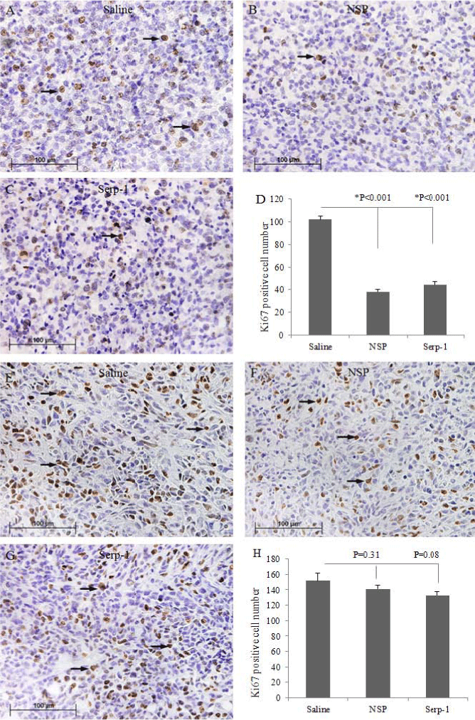
| Figure 3: Serp-1 treatment inhibits pancreatic cancer cell proliferation in vivo. Anti-Ki67 antibody was used to stain for proliferating cells (brown) in the tumor tissues. Pancreatic cancer Hs766t cell proliferation was inhibited in Serp- 1 and neuroserpin-treated mice. (A - Saline, B- NSP, C - Serp-1) Pancreatic cancer tissues isolated from NOD/SCID mice at 4 weeks follow up and stained for Ki67 (arrows point to representative dividing cells). (D) Bar graphs illustrate comparisons of mean ± SE for positively stained cell counts for Ki67 positive cells in pancreatic cancer sections. Ki67 positive cells in five randomly selected high power fields (HPFs) were counted for each tumor tissue. The averages of positive cell counts in each treatment group were compared using ANOVA (n=40 HPFs total, *: significant level of P< 0.05). Breast cancer cell proliferation was not inhibited in vivo by Serp-1 and NSP treatment. (E- Saline, F - NSP, G - Serp-1) Breast cancer with Ki67 staining (arrows point to representative dividing cells). (H) Bar graphs show comparison of Ki67 positive cells in breast cancer MD-231 cell implants after treatments (n=50 HPFs total). Magnification: 400X. |