 |
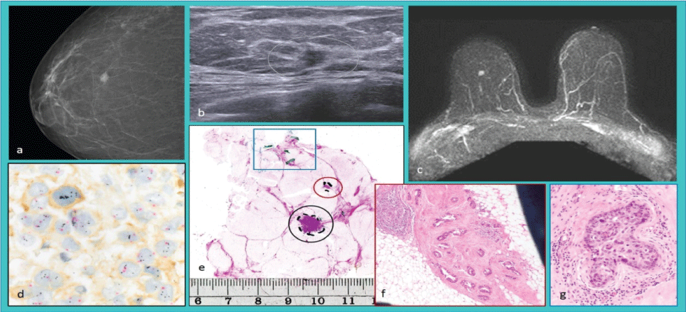
| Figure 2: Radiologicallyunifocal invasive breast carcinoma. (a) Radiogram, (b) Sonogram, and (c) Magnetic resonance image. (d) Tricolor bright field in situ hybridization showing HER2 gene amplification. (e) Large-format histology of the surgical specimen shows the radiologically detected invasive tumor focus (black circle), an additional invasive focus (red circle), and structures of the in situcarcinoma (blue box). (f) Magnification of the additional invasive focus and (g) in situ component stained with hematoxylin and eosin. |