|
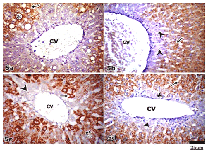
| Figure 5: Micrograph of immunoperoxidase technique for Hep Par-1 in both group II & III. (a): Shows heterogeneous granular cytoplasmic immunoreactivity (arrow) of hepatocytes with stronger expression around fatty vacuoles (asterisk). A wide area of negative immune expression (arrowhead) is seen around the central vein (cv), Subgroup IIA. (b): shows hepatocytes with moderate cytoplasmic immunoreactivity (arrow). A relatively wide area of negative immune reaction (arrowhead) is noticed around the central vein (cv). Subgroup IIB. (C): shows patchy immunoreactivity. Some hepatocytes showing strong immune reaction (arrow), others express weak reaction (double arrow). An irregular area of negative immune reaction (arrowhead) around the central vein (cv) is noticed. Subgroup IIIA. (d): shows moderate immunoreactivity in the cytoplasm of most hepatocytes (arrow) except a negative narrow zone (arrowhead) around the central vein (cv), Subgroup IIIB. |