 |
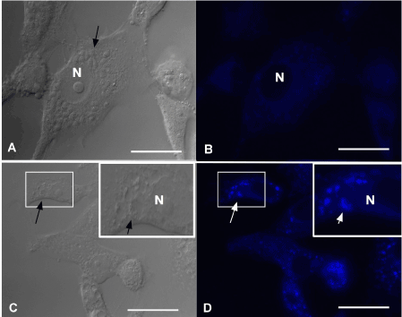
| Figure 6: Vero cells infected with amastigotes of T. cruzi for 36 h and stained with Monodansylcadaverine. Untreated cells (a and b) and treated with compound 1 at 1 mM for 12 h (c and d). Black arrows: amastigotes and white arrows: autophagic vacuoles containing parasites. N=nucleus. Scale bars: A,B=60 μm; C,D=100 μm. |