![]() |
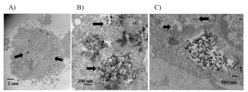
This figure shows that TiO2 nanoparticles were internalized by human skin fibroblasts shown by TEM imaging. The black arrows indicate TiO2 nanoparticle aggregates within different subcellular compartments. A. TEM image showing TiO2 nanoparticles located throughout the entire cell (4,000X). B. TEM image showing different sizes of TiO2 nanoparticle aggregates within the cytoplasm (10,000X). C. TEM image showing TiO2 nanoparticles within the nucleus (10,000X). |
Figure 4: TiO2 nanoparticles were internalized by human skin fibroblasts. |