 |
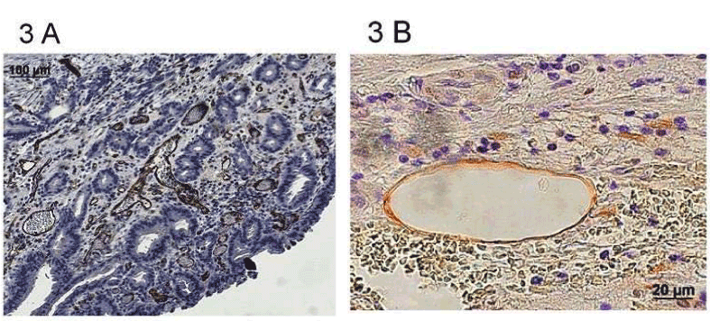
| Figure 3: Figure 3A shows a gastric cancer tissue sample from a T1m carcinoma after immunohistochemical staining against CD31 with enlarged pathological vessels (partially containing erythrocytes) in the neoplastic region. Figure 3B presents an immunohistochemical staining against VEGFR-2 with positive reaction of the vessel endothelium. |