 |
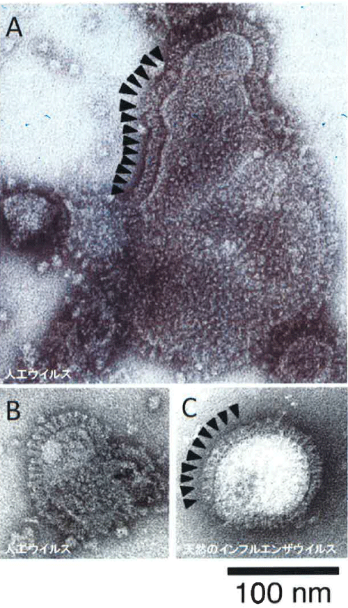
| Figure 8: Electron microscopy of purified VLP produced in silkworm pupae. A large VLP were observed in Figure 8A possessing about 300 nm in diameter and this was densely covered with influenza HA proteins. In contrast, VLP similar to authentic virus (B), this particle was very similar in size and structure to that of authentic virus (C). Arrows indicated the position of spikes. |