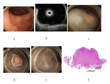
(a) A 6-mm-diameter carcinoid tumor in the upper rectum. (b) Thinning of the third layer by endoscopic ultrasonography. (c) Submucosal injection with hyaluronic acid. (d) Circumferential mucosal incision with a needle knife. (e) After the resection by snaring. (f) The vertical distance of the resection margin was negligible from the lower edge of the tumor.