 |
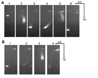
| Figure 8:Electrophoresis of plasmid pBR322 DNA treated with E. coli topoisomerases. (A) Panel 1, linear DNA marker (0.3 µg); Panel 2, supercoiled DNA marker (0.3 µg); Panel 3, supercoiled DNA treated with E. coli topoisomerase I (1U); Panel 4, supercoiled DNA treated with E. coli gyrase (1U); Panel 5, supercoiled DNA treated with E. coli topoisomerase III (1pmol); Panel 6, supercoiled DNA treated with E. coli topoisomerase IV (1U). (B) Panel 1, supercoiled DNA (0.3 µg) relaxed by E. coli topoisomerase I (1U); panel 2, the DNA sample of panel 1 (0.3 µg) treated with E. coli gyrase (1U); panel 3, supercoiled DNA (0.3 µg); panel 4, supercoiled DNA (2 µg) treated with E. coli topoisomerase IV(1U). The first dimension (with 3 µg/ml of chloroquine) was from top to bottom and the second dimension (with 15 µg/ml of chloroquine) was from right to left. |