|
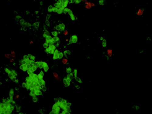
| Figure 1: Positive result of DFA technique seen under a fluorescent microscope for the detection of hMPV antigen in nasopharyngeal swab using FITC stain. The psuedostratified columnar epithelial cells of the nasopharynx, shows prominent green fluorescence in cytoplasm (arrow), while the nuclear region is pale. |