|
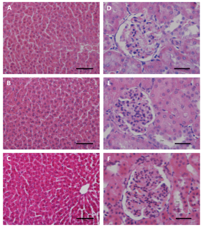
| Figure 5: Histological study of liver and kidney. Staining with hematoxylin (cell nuclei-blue) and eosin (cytoplasm-pink/red). A-C: Microscopic analysis of liver: Negative control group (A); Animals treated with IBU-loaded PLA nanoparticles (B); Animals treated with free IBU (C); 20x magnification in the original, Bar=50 μm. D-F: Microscopic analysis of kidney: Negative control group (D); Animals treated with IBU-loaded PLA nanoparticles (E); Animals treated with free IBU (F). 40X magnification in the original, Bar=25 μm. |