

 |
 |
 |
 |
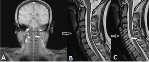
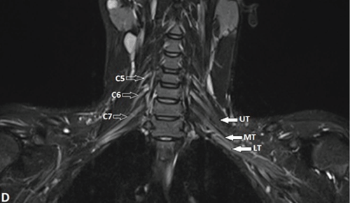
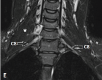
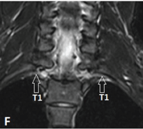
| Figure 1: Normal MR imaging planes and appearance of BP. A-F, Acquisition of the coronal plane with fat-suppressed T2-weighted anterior (D), mid (E), andposterior (F) coronal sections. First a coronal cervical spine scout is obtained (A), based on this a sagittal T2-weighted spin-echo sequence of the cervical spine isacquired (B). Now based on this sagittal image, coronal fat-suppressed T2-weighted and T1-weighted sequences are obtained in a plane parallel to the long axis ofthe lower cervical vertebrae (C) because the BP is oriented in an oblique coronal plane. On coronal fat-suppressed T2-weighted sequence, long axis of the nerveplexus is seen as elongated, uniform calibre, mildly hyperintense fascicular structure. On the anterior coronal image, normal C5, C6, C7 nerve roots (open arrows)are seen on the right side; on the left, the upper (UT), middle (MT) and lower trunks (LT) (solid arrows) are seen (D). At the level of middle scalene muscle (star), onthe mid-section, C8 ventral ramus (open arrows) is seen bilaterally (E), while the T1 nerve root (open arrows) is visualized completely on the posterior section (F). | |