|
||
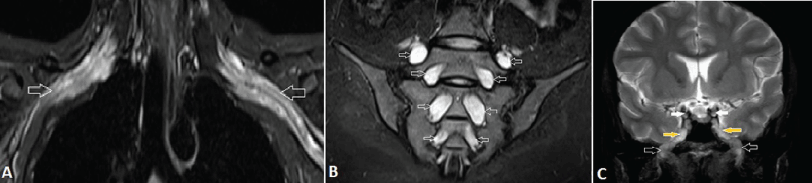
| Figure 12: A 29-year-old female with chronic inflammatory demyelinating polyneuropathy (CIDP). A, Coronal fat-suppressed T2-weighted MR image shows bilateral symmetrically enlarged and hyperintense BP (arrows). B, Coronal fat-suppressed T2-weighted MR image shows diffuse marked symmetrical hypertrophy and abnormal high signal of the lumbosacral nerve roots (arrows). C, Coronal T2-weighted MR image of brain shows marked thickening of the cranial nerves in the lateral wall of cavernous sinus and foramen ovale. Note the enlarged oculomotor nerves (white arrows) and mandibular nerves (black arrows), coming out from Meckel’s cave (yellow arrows) into the masticator space through the enlarged foramen ovale. | ||