|
||
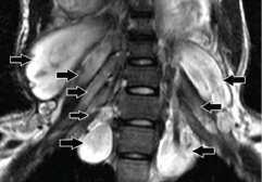
| Figure 14: A 16-year-old male with neurofibromatosis type 1. Coronal fatsuppressed T2-weighted MR image shows diffuse, fusiform, longitudinally oriented hyperintense lesions involving all the pre and postganglionic cervical nerve roots (arrows) of BP bilaterally without any definite capsule. | ||