|
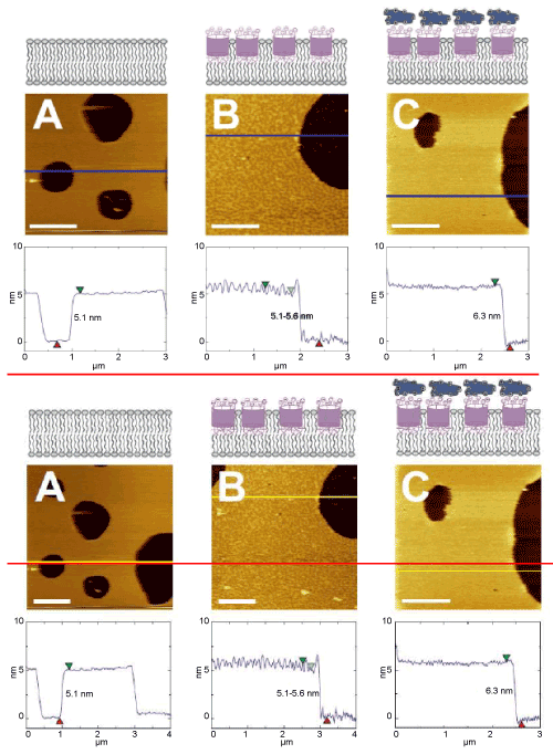
| Figure 1: Topographical investigation of the DMPC self-assembled monolayer performing AFM imaging. (A) Bare DMPC bilayer on mica. The height difference between bilayer and mica is 0.5 nm. The RMS of the surface roughness on the layer is 0.18 nm. (B) DMPC layer with embedded Calix[6]arene molecules. Two phases are visible. The lower phases have a height of 5.1 nm whereas the higher areas have a height of 5.6 nm. The RMS of the surface roughness on the layer is 0.49 nm. (C) DMPC layer with embedded Calix[6]arene molecules after the addition of Cytochrome c. The height difference increased to 6.3 nm and the surface roughness had a RMS of 0.32 nm without detectable phase separation. z-Scale for all images is 12 nm. |