|
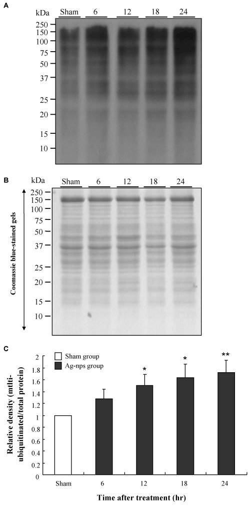
| Figure 2: Changes in mitochondrial ubiquitinated proteins expression in liver tissues at various time points following i.p. injection of Ag-nps. A: Mitochondrial ubiquitinated proteins expression based on Western blot analysis. B: Coomassie blue-stained gels demonstrating equal loading of the samples. C: The relative densities were normalized from part A to the total amount of protein measured from part B. The experiments were performed as described under Material and Methods. Empty columns represent sham groups while filled columns represent Ag-nps treatment groups. Molecular weight size markers (kDa) are indicated at the left. Values were means ± SD (n=8), *p<0.05 and **p<0.01 compared to sham group. |