|
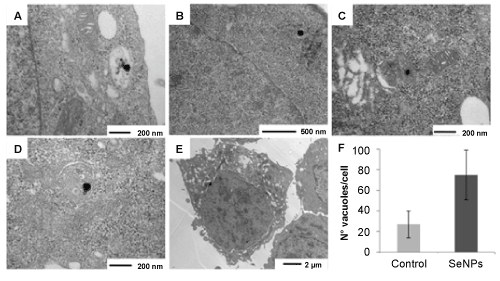
| Figure 3: Transmission electron microscopy showed that Ch-SeNPs uptaked by HepG2 cells are localized in (A) vacuoles, (B) throughout the cytoplasm and in subcellular organelles such as (C) mitochondria and (D) the endoplasmic reticulum. As expected, Ch-SeNPs were not localized in the nuclei due to their particle size. (E, F) About 90% of cells exposed to Ch-SeNPs exhibit more than 2-fold increased in the number of vacuoles as compared to control cells. |