|
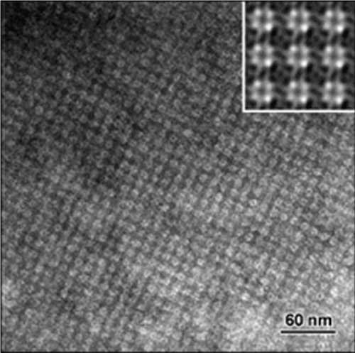
| Figure 3: Bacterial S-layer of Bacillus sphaericus JG-A12 shown under Transmission Electron Microscopy. The picture illustrates the symmetry of S-layers and allows the visualisation of the pores where metallic nanoparticles are localised. Reprinted from Pollman et al., with permission from Elsevier: Biotechnology Advances, copyright 2005 [36]. |