|
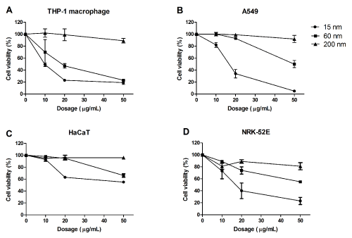
| Figure 2: Cytotoxicity of the SiO2 NP in four different cell lines. Cells were treated with SiO2 NP at 0, 10, 20 and 50 μg/mL for 24h. Cell viabilities were determined using WST-1 assays and expressed as percentage of untreated control cells. Experiment was repeated in 3 independent runs with using 4 replicas each. Data are given as means ± SEM for THP-1 macrophages (A), A549 (B), NRK-52E (C) and HaCaT cells (D). |