 |
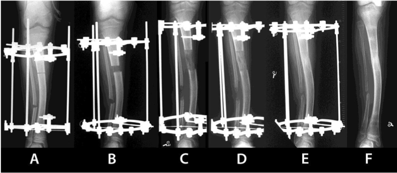
| Figure 3: Radiological representation of the distraction osteogenesis technique. A. Osteotomy is performed after the application of the external fixator; B. Initial phase of the distraction procedure. C. Elongation of the bone; D. New bone tissue formed at the distraction site; E. Radiological evidence of bone mineralization during the consolidation phase; F. Final consolidation of the lengthened bone and removal of the fixator. |