|
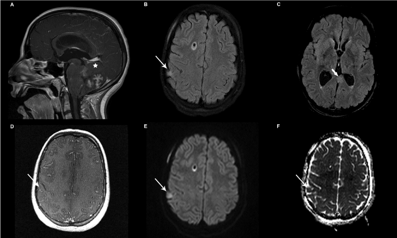
| Figure 1: Images from the MRI study at presentation showing the enhancing posterior fossa medulloblastoma (A- white asterisk). Axial FLAIR images demonstrating the hyperintense right parietal lesion considered a parenchymal metastatic deposit (B-thin white arrow) and left medial temporal lesion (C–thick white arrow). No enhancement is seen in the right parietal lesion (D-thin white arrow). B=1000 diffusion image (E) and corresponding ADC map (F) demonstrating restricted diffusion in the right parietal lesion (thin white arrow). |