|
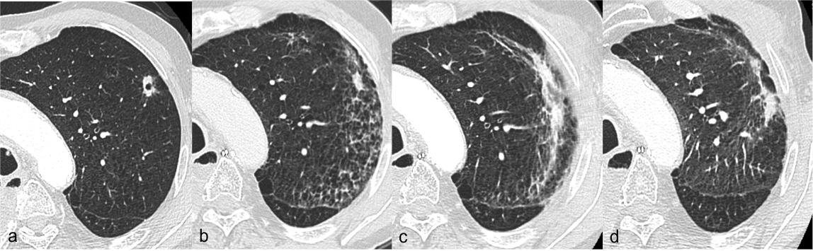
| Figure 1: Radiation pneumonitis (grade 1) radiological evolution. Case 6 Supplementary Table 4; a: CT scan at 1 month after the end of T-SBRT showing the pulmonary lesion; b: CT scan at 5 Months with the development of ground-glass opacities; c: CT scan at 9 months showing the maximal extension of infiltrates and appearance of solid opacities; d: CT scan at 13 months showing the opacity regression. |