|
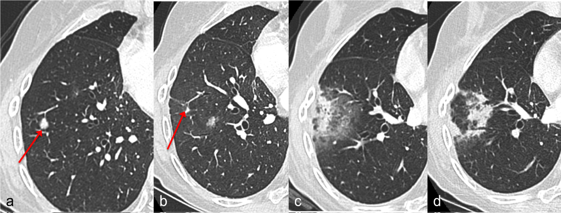
| Figure 2: Organising pneumonia radiological evolution. Case 9 Supplementary Table 4; a: CT scan at 2 months before the start of T-SBRT, the red arrow indicate one of the two irradiated lesions; b: CT scan at 4 months after the T-SBRT, the red arrow shows a decrease in the volume of the lesion; c: CT scan at 6 months (maximal extension of infiltrates) showing solid and ground-glass peripheral opacities which prevented the recognition of the malignant lesion; d: CT at 9 months showing the substantial resolution of previous solid infiltrates and the developing of others solid infiltrates in place of previous ground-glass opacities. |