|
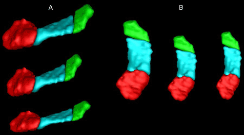
| Figure 4: Temporal order of events in incomplete HC “rebound” after traumatic brain injury. Compared to a control HC (A from lateral and B from superior, top and left), the HC in acute period after a TBI is projected anteriorly and squashed (middle). Over the next 12 to 24 months, the HC stretches back towards its original length but never fully rebounds as it becomes thinner as represented by less medial and lateral volume (A bottom, B far right). Red=head, blue=body, green=tail. Note that the division between the tail and body, which represents the fornix, appears to shift forward and then backwards over time. Note this is a simulated series of images; that is, they have been artificially stretched in order to more clearly illustrate the HC rebound principle. |