|
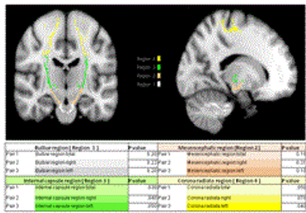
| Figure 1: MRI coronal (left) and sagittal view (right). Fractional anisotropy of the PT was analyzed, measured and compared in 4 regions of interest (ROI) named as bulbar (ROI-1, in white), mescencephalic (ROI-2, brown), posterior limb of internal capsule (ROI-3, green)) and Corona Radiata (ROI-4, yellow). Differences between baseline and after stem cells transplantation MRI-FA are shown. Only ROI 3 and 4 that are located close to the site of transplantation showed statistically significant increment in FA in comparison to baseline FA. In the corona radiata, increase in FA was found when left and right sides were measured and compared in total (p=0.05), FA in this ROI showed statistically significant increment only in left side(p= 04). |