|
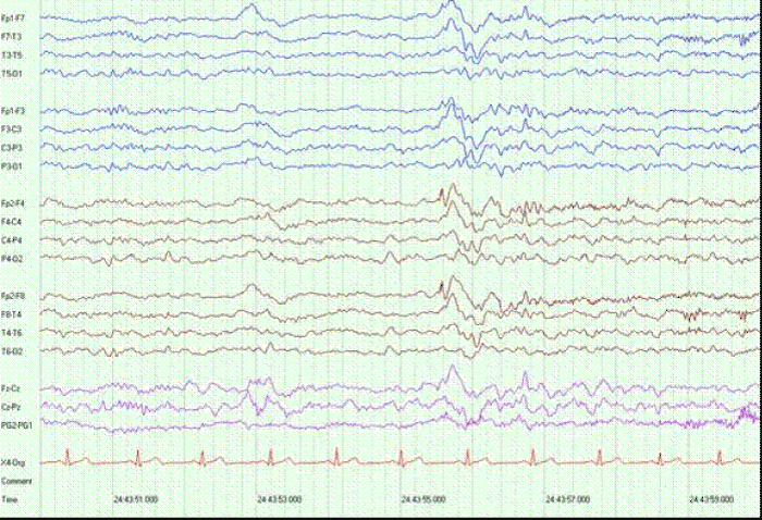
| Figure 4: Ten-second page of EEG from patient 5, demonstrating an abnormally shaped K complex. On the top panel, a sharp component is seen with a likely frontal location.Bottom panel demonstrates the same abnormality on transverse montage. The phase reversal appears at FP2. |