|
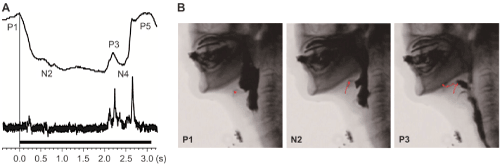
| Figure 4: Interruption of swallowing in a 72-year-old woman with Parkinson’s disease. (A) The swallowing waveform shown by the magnetic swallowing monitoring device. After the start of swallowing, the interval between P1 and P3 was approximately 2.2 s. The sound waveform nearly agreed with P3. The upper waveform represents the distance waveform, and the lower waveform represents the sound waveform. (B) On videofluorography, after the swallowing reflex had started (P1), the movement of the hyoid bone (red line) stopped with the larynx still elevated (N2). The hyoid bone then moved anteriorly, and the cricopharyngeal muscle opened. The liquid passed through the pharynx (P3). |