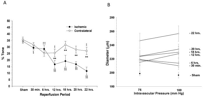
(A) Graph showing percent tone of MCA exposed to different periods of reperfusion all with 30 minutes of ischemia compared with sham-operated control animals. Shown are responses of MCA from both ipsilateral (closed circles) and contralateral (open circles) to occlusion. The amount of tone significantly diminished compared with sham-operated control in both contralateral and ischemic arteries as reperfusion duration increased. **P<0.01 contralateral versus ischemic; ‡P<0.05 contralateral versus sham control; #P<0.05 contralateral versus sham control; §P<0.05 ischemic versus sham control: §§P<0.01 ischemic versus sham control.
(B) Graph showing diameter of MCA after step increases in transmural pressure from 75 to 125 mm Hg after exposure to different periods of reperfusion all with 30 minutes of ischemia. Myogenic reactivity of MCA significantly diminished as reperfusion duration increased. From Cipolla MJ et al., Stroke 2002; 33: 2094-2099.