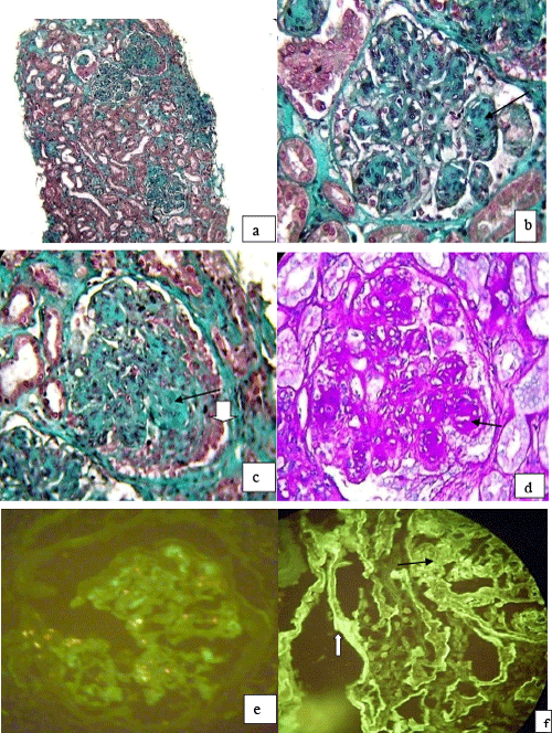
(a) Masson trichrome x 200. (b) Nodular glomerulosclerosis (c)
Nodular glomerulosclerosis (black arrow) and increasing fiber-cell (white arrow). Masson trichrome x 400. (d) PAS-positive nodule. NOT x 400. - Immunofluorescence:
(e) Absence of λ chain deposits (f) deposits κ chains in the glomerulus (black arrow) and tubular basement membranes (white arrow).