|
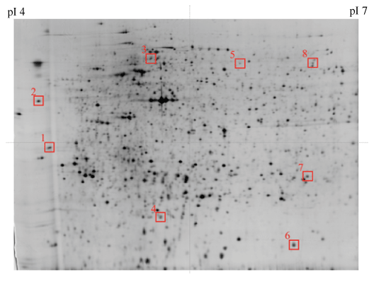
| Figure 2: Representative image of gels from benzene metabolites exposure (control). Fifty micrograms of proteins were applied to a pH 3-7 IPG strip (24cm), and with 12.5% constant vertical SDS-PAGE as the second dimension. The gel was visualized by silver staining, and the resulting image was analyzed by PDQuest software. Marked squares show specifi c spots to exposure. |