|
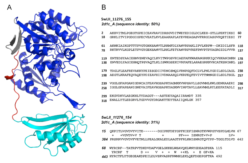
| Figure 3: Domain fusion model created for proteins 11276_155 and 11276_154, compared with T. thermophilus isocitrate dehydrogenase. (A) Structure of T. thermophilus isocitrate dehydrogenase (PDB 2d1c) is colored by its correspondence to the Leptospirillum Group II isocitrate dehydrogenase proteins: 11276_155, royal blue; 11276_154, cyan; N-terminal and linker regions with no corresponding residues in 11276_155, grey; and, linker region with very poor alignment to N-terminal region of 11276_154, red. (B) Sequence alignments calculated between PDB template 2d1c chain A and proteins: 11276_155 (top), and 11276_154 (bottom). |